What Is Mass Spectrometry in Peptide Identity Testing?
What Is Mass Spectrometry in Peptide Identity Testing?
RESEARCH USE ONLY
This guide explains analytical interpretation of mass-spectrometry evidence for laboratory research materials. It does not provide dosing, administration, reconstitution, treatment or veterinary-use guidance.
What does mass spectrometry confirm in peptide testing?
Mass spectrometry (MS) measures ions according to their mass-to-charge ratio (m/z). For a synthetic peptide whose intended sequence and chemical form are already defined, intact-mass MS can provide strong identity evidence when the observed ion pattern or deconvoluted mass is consistent with the theoretical mass of that exact peptide form.
The crucial word is ‘consistent’. A mass match is not the same as complete structural proof. Different structures can sometimes share the same or effectively indistinguishable mass, and an MS result does not replace chromatographic purity evidence. For that reason, peptide quality documentation is stronger when mass-based identity evidence is interpreted alongside HPLC or another appropriate separation method.
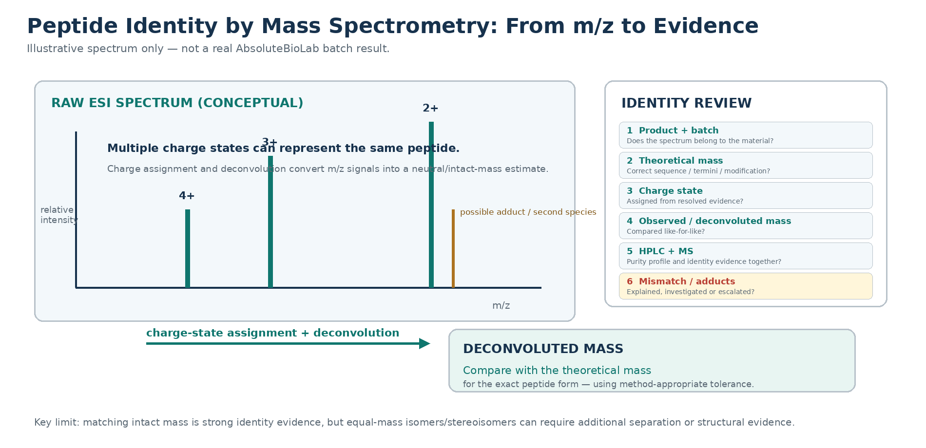
Mass-to-charge ratio is not the same as neutral molecular mass
A mass spectrometer does not directly display the neutral peptide molecular mass on the raw x-axis. It records m/z values for ions. In electrospray ionisation (ESI), peptides commonly acquire more than one charge, so the same peptide can appear as several peaks or isotope envelopes at different m/z values.
Electrospray’s ability to produce multiply charged ions is one of the developments that made high-mass biomolecules accessible to mass analysis. Early high-resolution ESI work showed that one molecular species can generate multiple charge states and that isotope spacing can be used to assign charge.[3]
| Term | Meaning | Common interpretation error |
|---|---|---|
| m/z | Mass-to-charge ratio of an ion detected by the instrument. | Reading the raw m/z as if it were the neutral peptide mass. |
| Charge state (z) | Number of elementary charges carried by the ion. | Assuming a charge state without spectral evidence. |
| Isotope spacing | Spacing among isotopic peaks within a resolved ion envelope; can support charge assignment. | Treating a noisy or unresolved envelope as definitive. |
| Deconvoluted mass | Mass estimate reconstructed from one or more charge-state signals. | Assuming software output is independent of charge/adduct assignments or processing settings. |
| Theoretical mass | Calculated mass for the exact sequence and defined chemical form. | Using a theoretical value for the wrong termini, modification or peptide form. |
How charge states are assigned
For sufficiently resolved spectra, isotopic spacing provides direct charge-state information: as charge increases, isotopic peaks within the envelope are separated by a smaller m/z interval. Multiple charge states of the same analyte should also reconstruct to a consistent neutral mass when correctly assigned.
Charge-state assignment should not be reduced to a rule such as ‘small peptides are always 1+ or 2+’. Ionisation depends on sequence, basic sites, solvent conditions, instrument settings and other factors. The current live page’s statement that [M+H]+ and [M+2H]2+ are the most common ions for essentially all 500-5000 Da research peptides should therefore not be retained as a universal rule.
Compare observed and theoretical mass on the same basis
A meaningful mass comparison requires like-for-like values. Before deciding that a result matches, check how both numbers were defined.
This is also why the article should not publish a universal ‘+/-0.1 Da confirms identity’ rule. Appropriate mass error depends on instrument resolving power, calibration, acquisition mode, isotope assignment, analyte mass, the value being compared and the laboratory’s validated or fit-for-purpose procedure. ICH Q2(R2) frames analytical performance in terms of the intended purpose of the procedure rather than one universal tolerance.[1]
| Question | Why it matters |
|---|---|
| Is the theoretical mass monoisotopic or average? | A monoisotopic exact mass and an average molecular mass are different quantities and should not be compared as though they were identical. |
| Does the theoretical value include the correct termini? | Amidation, acetylation and other terminal forms change the elemental composition and mass. |
| Are deliberate modifications included? | Labels, lipidation, PEG-like groups, linkers and other modifications alter the expected mass. |
| Is a disulfide or other covalent state relevant? | Bond formation or chemical modification can change the expected mass; interpretation must match the defined structure. |
| Is the reported value raw m/z or a deconvoluted neutral mass? | A raw ion m/z cannot be compared directly with a neutral theoretical mass without the correct charge/adduct model. |
Adducts and secondary ion series can complicate interpretation
Peptide spectra can contain more than the protonated molecular ion series. Alkali-metal adducts, solvent-related species, in-source fragments, oxidation/dehydration products or other sample components can produce additional signals. A second peak series should therefore be interpreted from its charge-state pattern, exact mass relationship, chromatographic behaviour and method context rather than labelled automatically from a single mass shift.
High-resolution MS studies have specifically shown that adduct and impurity ions can affect mass interpretation when the spectral envelope is not resolved or correctly assigned.[3] If an unexpected series is material to the identity decision, it should be explained, investigated or escalated rather than hidden by deconvolution settings.
What intact MS can and cannot establish
| Intact MS can support | Intact MS cannot establish by itself |
|---|---|
| Consistency of observed/deconvoluted mass with the intended peptide form. | Chromatographic purity or the amount of peptide in the vial. |
| Detection of some species whose mass differs from the target. | The structure of every unexpected species from mass shift alone. |
| Recognition of multiple charge states and some adduct patterns. | Sterility, endotoxin, biological activity or long-term stability. |
| Evidence that the dominant detected species has the expected mass. | Amino-acid sequence in all cases without fragmentation or other structural evidence. |
| Detection of some mass-changing modifications or truncations. | Stereochemistry; D- and L-configurations have the same elemental mass. |
| Exact-mass discrimination between some closely related compositions when resolution is sufficient. | Distinction between all isobaric or isomeric structures. |
Why equal mass does not always mean equal structure
This is one of the most important limits in peptide identity testing. Two different structures can share the same elemental composition or have masses that cannot be distinguished by a particular intact-MS experiment.
Practical peptide-synthesis work has demonstrated this problem. In an orthogonal CE-UV-ES-MS study, synthetic-peptide components with the same nominal molecular mass were observed despite evidence that more than one component was present.[4] High-resolution peptide MS studies have also differentiated peptides with identical monoisotopic masses only after collision-induced fragmentation supplied sequence-related information.[5]
Stereoisomers are an even clearer example: D- and L-amino-acid configurations have the same mass. Chiral separation coupled with MS/MS can be needed when stereochemical purity is the analytical question. Therefore, a correct intact mass is strong identity evidence, but it should not be described as proof that every residue has the intended stereochemistry or sequence arrangement.
What MS/MS adds to intact-mass evidence
Tandem mass spectrometry (MS/MS) isolates a precursor ion and fragments it. The resulting product-ion pattern can provide sequence-related structural evidence because peptide-bond fragmentation generates ions that reflect parts of the peptide backbone.
For a known synthetic peptide, MS/MS can therefore increase confidence beyond a simple intact-mass match, particularly when distinguishing certain sequence variants or locating modifications. It is still not a universal solution: some isomeric or stereochemical differences require specialised fragmentation, chromatographic separation, chiral methods or another orthogonal technique.
The analytical question should determine the evidence required. An intact-mass check may be sufficient for one laboratory decision, while another method may require sequence-level or stereochemical confirmation.
ESI-MS and MALDI-MS are different ionisation workflows
Electrospray ionisation (ESI) and matrix-assisted laser desorption/ionisation (MALDI) are both established ways to introduce peptides into a mass spectrometer, but they produce different spectral behaviour and fit different workflows.
Neither ionisation method should be treated as intrinsically ‘acceptable’ or ‘unacceptable’ without reference to the purpose and performance of the actual method. The analytical procedure must be fit for the identity question being asked.
| Feature | ESI / LC-MS | MALDI-TOF-MS |
|---|---|---|
| Ionisation behaviour | Often produces multiple charge states for peptides. | Often dominated by singly charged ions for many peptide applications, though spectral behaviour is method-dependent. |
| Coupling to separation | Readily coupled online to liquid chromatography. | Commonly analysed from a prepared target after mixing with a matrix. |
| Strength for peptide documentation | Can combine chromatographic retention/profile information with mass detection. | Can provide rapid mass profiling and identity support for suitable samples. |
| Interpretation focus | Charge-state assignment, adducts and deconvolution are central. | Matrix/background effects, calibration and peak assignment are important. |
Why HPLC plus MS is stronger than either result alone
HPLC and MS answer complementary questions. HPLC can separate detected components and provide a chromatographic purity/profile result; MS can provide molecular-mass and identity evidence for detected ions. A main HPLC peak without specific identity evidence may be the wrong compound, while an intact MS mass match does not show what fraction of the sample is represented by that species.
ICH Q6A makes the same general specificity point in its regulated pharmaceutical context: identification by a single chromatographic retention time is not sufficiently specific, while combined procedures such as HPLC/MS can provide stronger identification evidence.[2] The guidance is cited for the analytical principle, not as a regulatory requirement for Core Research RUO products.
HPLC interpretation: What Is HPLC Purity in Peptide Testing?
Full CoA interpretation: How to Read a Peptide Certificate of Analysis
| Evidence | Primary question | Key limitation |
|---|---|---|
| HPLC-UV purity/profile | How much included detector area is assigned to the main chromatographic peak? | Does not prove molecular identity by retention time alone. |
| Intact MS | Is detected mass consistent with the defined peptide form? | Does not provide chromatographic purity and may not distinguish isomers. |
| LC-MS | How do chromatographic components relate to mass signals? | Still depends on separation, ionisation, detector response and structural specificity. |
| MS/MS | Do fragment ions support sequence-related structure? | Not all isomeric/stereochemical questions are resolved. |
From Our Work: how Core Research reviews peptide MS evidence
When reviewing mass-spectrometry evidence on a peptide CoA, Core Research checks five things: (1) observed molecular mass against the theoretical mass; (2) the stated charge state or deconvoluted mass; (3) whether the MS result belongs to the correct product and batch; (4) whether HPLC purity and MS identity evidence are both present where that documentation is being relied upon; and (5) whether any meaningful mass mismatch requires clarification. This describes the supplied document-review workflow only; it does not imply that Core Research performed the laboratory analysis unless the batch record explicitly states that.
What should be visible on a useful peptide MS record?
| Field / evidence | Why it matters | Red flag |
|---|---|---|
| Product / exact entity and batch | Connects the spectrum or report to the received material. | MS result cannot be matched to the batch. |
| Ionisation / method type | Provides context for expected ion and charge behaviour. | Only “MS confirmed” is stated with no method or data. |
| Theoretical mass basis | Shows what chemical form and mass definition are being compared. | Theoretical value does not match the labelled peptide form. |
| Observed m/z and charge state, or deconvoluted mass | Makes the identity comparison independently interpretable. | A bare pass/fail statement with no observed value. |
| Spectrum or suitable analytical output | Allows review of ion series, secondary signals and overall context where provided. | Only a single number is shown without enough context to interpret it. |
| Mass error / acceptance logic appropriate to the method | Shows how the laboratory judged agreement. | A universal tolerance is asserted with no instrument/method basis. |
| Orthogonal HPLC or other evidence where required | Separates identity and purity questions. | One method is presented as proving every quality attribute. |
Frequently asked questions
Does a matching molecular mass prove the peptide sequence?
It provides strong evidence that the detected species has the expected mass, but equal-mass isomers or stereoisomers can exist. Sequence-level confidence may require MS/MS or another orthogonal method depending on the question.
What is a charge state in peptide mass spectrometry?
It is the number of charges carried by the detected ion. In ESI, one peptide can appear at several charge states, giving several m/z peaks that deconvolute to the same neutral mass when correctly assigned.
Why is a deconvoluted mass different from the raw m/z value?
The raw spectrum records mass-to-charge ratio. Deconvolution uses the assigned charge-state pattern to estimate the underlying neutral/intact mass.
Is +/-0.1 Da the universal acceptance limit for peptide identity?
No. Appropriate mass error depends on the instrument, calibration, resolving power, analyte mass, isotope/adduct assignment and method purpose. The acceptance rule should come from the fit-for-purpose analytical procedure.
Can mass spectrometry measure peptide purity?
MS can detect and characterise many impurity species, especially when coupled to chromatography, but a simple intact-mass match is not a chromatographic purity measurement. HPLC and MS should not be treated as interchangeable tests.
Can MS distinguish D- and L-amino-acid versions of a peptide?
Not from intact mass alone because stereoisomers have the same elemental mass. Chiral separation or other stereochemistry-sensitive methods may be required.
Key takeaway
Peptide mass spectrometry is strongest when read as an identity-evidence chain: confirm the exact product and batch, understand whether the reported value is m/z or deconvoluted mass, assign charge states correctly, compare observed and theoretical mass on the same basis, inspect unexplained secondary signals, and combine the result with HPLC or other orthogonal evidence where the quality decision requires it. A matching mass is powerful evidence, but it is not chromatographic purity, stereochemical proof or a universal guarantee of structure.
References
- ICH Q2(R2). Validation of Analytical Procedures. Final guidance, March 2024. Used for fit-for-purpose analytical-performance principles; not presented as a regulatory requirement for Core Research RUO materials.
- ICH Q6A. Specifications: Test Procedures and Acceptance Criteria for New Drug Substances and New Drug Products: Chemical Substances. Used for the analytical specificity principle and complementary identification methods; not presented as an RUO regulatory requirement.
- McLafferty FW, Kelleher NL, Begley TP, Fridriksson EK, Zubarev RA, Horn DM. High-resolution tandem mass spectrometry of large biomolecules. Proceedings of the National Academy of Sciences USA. 1992;89(1):286-290. DOI: 10.1073/pnas.89.1.286.
- Rosnack KJ, Stroh JG, Singleton DH, Guarino BC, Andrews GC. Use of capillary electrophoresis-electrospray ionization mass spectrometry in the analysis of synthetic peptides. Journal of Chromatography A. 1994;675(1-2):219-225. DOI: 10.1016/0021-9673(94)85275-8.
- Huddleston MJ, Bean MF, Carr SA. High resolution electrospray mass spectrometry with a magnetic sector instrument: accurate mass measurement and peptide sequencing. Rapid Communications in Mass Spectrometry. 1995;9(12):1044-1051. PMID: 7612937.
- Liu Y, Gong X, et al. Enantiomeric purity analysis of synthetic peptide therapeutics by direct chiral high-performance liquid chromatography-electrospray ionization tandem mass spectrometry. Journal of Chromatography B. 2023;1219:123653. PMID: 36857849. Used to illustrate the need for stereochemistry-sensitive separation when equal-mass D/L impurities are the question.